Vol. 7, Issue 1 (2019)
Glutaraldehyde and Osmium tetroxide fixation of thymus in effective demonstration of Hassal’s corpuscles in thymus of goats
Author(s): A Kumaravel, S Sivagnanam and S Paramasivan
Abstract: The scanning electron micrograph of the goat thymus was studied by glutaraldehyde and osmium tetroxide fixation. The thymus showed epithelial reticular cells, thymocytes, macrophages, Hassal’s corpuscles, interdigitating cells, Dendritic cells and Thymic nursing cells. The epithelial cells form a meshwork in the thymus parenchyma. Cortical epithelial reticular cells were stellate in shape, while the medullary epithelial reticular cells were of two types, stellate and large vacuolated elements. A continuous single layer of epithelial cells separates the parenchyma from connective tissue formations of the capsule, septa and vessels. Surrounding the blood vessels, this epithelial sheath was continuous in the cortex, while it was partly interrupted in the medulla, suggesting that the blood thymus barrier might function more completely in the cortex. Osmium tetroxide being a heavy metallic compound gives higher electron beam resistance under the gold sputtered surface of the tissue.
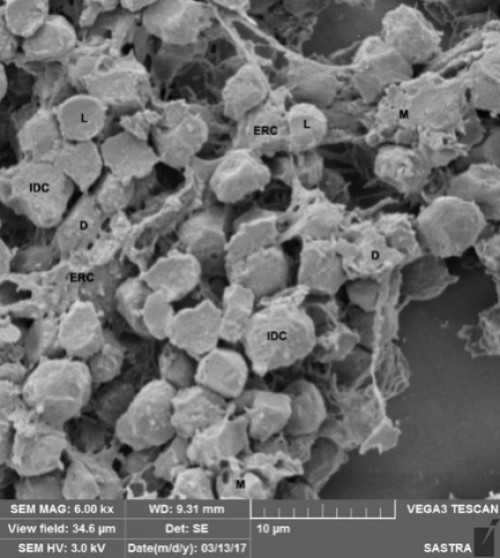
Fig. 1: L – Lymphocyte, M – Macrophage, D – Dentritic Cell, ERC – Epithelial Reticular Cell and IDC – Inter Digitating Cell
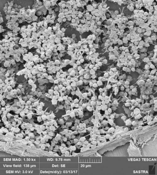
Fig. 2: IDC – Inter Digitating Cell, L – Lymphocyte, ERC – Epithelial Reticular Cell and C – Capsule
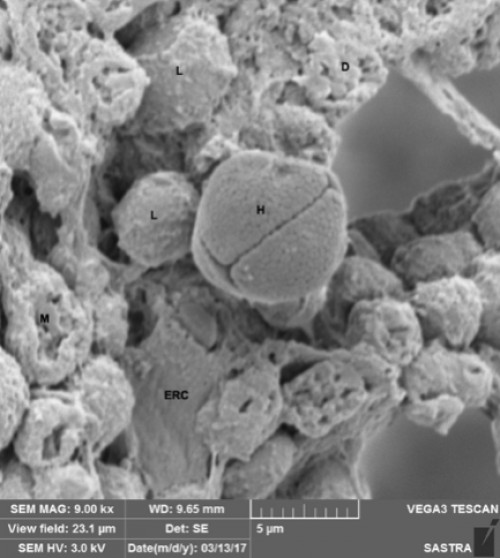
Fig. 3: L – Lymphocyte, D – Dendritic Cell, H – Hassel’s Corpuscle, M – Macrophage and ERC – Epithelial Reticular Cells.
Pages: 141-143 | 814 Views 54 Downloads
download (7267KB)


How to cite this article:
A Kumaravel, S Sivagnanam, S Paramasivan. Glutaraldehyde and Osmium tetroxide fixation of thymus in effective demonstration of Hassal’s corpuscles in thymus of goats. Int J Chem Stud 2019;7(1):141-143.