Vol. 7, Issue 1 (2019)
Scanning electron microscopy of paraffin embedded white pulp of spleen of goats
Author(s): A Kumaravel, S Sivagnanam and S Paramasivan
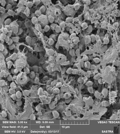
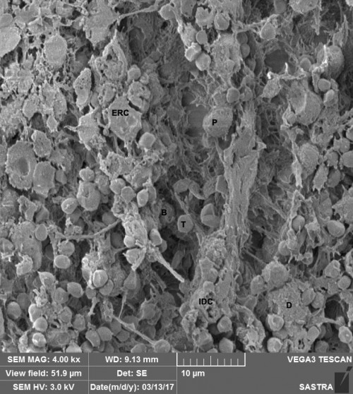
Abstract: The paraffin embedded tissue sections and the cryofractured tissue obtained by conventional processing for electron microscopy are compared under scanning electron microscope. The routine processing of white pulp of the spleen procured from the slaughter of goats by paraffin embedding method revealed better results than conventional processing for electron microscopy. Epithelial reticular cells, T lymphocytes, B lymphocytes, plasma cells, dentritic cells and interdigitating cells were observed in both methods at scanning electron microscope. The clarity and contrast of depth of focus was better at magnification of 4000 in deparaffinized specimen. The paraffin processing method was simple and less expensive and combines the advantages of great depth of focus and high resolution of scanning electron microscope with the simple preparatory techniques employed for light microscopy than the conventional electron microscope processing techniques which is very expensive.

Fig. 1: Scanning electron micrograph of the white pulp of spleen in 2 year old goat processed by conventional EM method. T – T lymphocyte, B – B lymphocyte, P – plasma cell, D – Dentritic cell, IDC – Interdigitating cell and ERC – Epithelial reticular cell.

Fig. 2: Scanning electron micrograph of the white pulp of spleen in 2 year old goat processed by Paraffin embedding method. T – T lymphocyte, B – B lymphocyte, P – plasma cell, D – Dentritic cell, IDC – Interdigitating cell and ERC – Epithelial reticular cell.
Pages: 26-28 | 1592 Views 117 Downloads
download (7457KB)
How to cite this article:
A Kumaravel, S Sivagnanam, S Paramasivan. Scanning electron microscopy of paraffin embedded white pulp of spleen of goats. Int J Chem Stud 2019;7(1):26-28.


